
CBCT vs OPG for TMJ Diagnosis in Hyderabad: Why CBCT May Be Recommended for Jaw Joint Evaluation
If you’re looking for TMJ treatment in Hyderabad because your jaw clicks every time you chew or you wake up with a dull ache near your ear, you’ve probably searched for answers online more than once. One question that comes up repeatedly from patients across Hyderabad is simple but important: should you get an OPG or a CBCT scan for TMJ diagnosis?
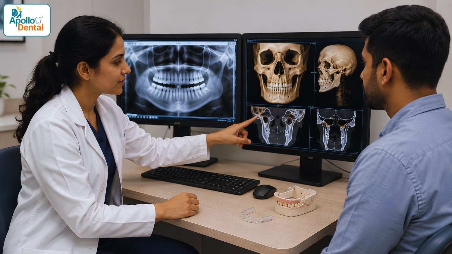
Both are dental X-rays. Both can be done in a single sitting. But they don’t show your jaw joint in the same way, and picking the wrong one can mean a missed diagnosis. At Apollo Dental Nallakunta, patients dealing with jaw pain, clicking sounds, or restricted mouth opening are guided through this decision by Dr. Ancy V. Ignatius, a TMD (temporomandibular disorder) specialist with over 17 years of experience in oral medicine and radiology. This article breaks down what each scan does, why CBCT is often the better fit for TMJ cases, and what to expect if you’re getting evaluated in Hyderabad.
What Is an OPG Scan?
An OPG (Orthopantomogram), sometimes called a panoramic X-ray, is a single flat image that captures your entire jaw, teeth, and sinuses in one wide sweep. It’s quick, affordable, and one of the most used dental screening tools.
For general dental checks, OPG works well. It helps dentists spot:
- Cavities and impacted wisdom teeth
- Bone loss around teeth
- Cysts, tumors, or other jaw abnormalities
- A rough overview of jaw joint alignment
However, an OPG produces a 2D image, it flattens a 3D structure onto a single plane. The temporomandibular joint (TMJ) is a small, curved, and layered structure, and on an OPG it often overlaps with nearby bones. This overlap can hide fine cracks, early bone erosion, joint space evaluation or subtle changes in the joint that matter most when diagnosing TMJ disorders.
What Is a CBCT Scan?
CBCT (Cone Beam Computed Tomography) is a more advanced imaging technique that captures hundreds of thin cross-sectional images and reconstructs them into a detailed 3D model of your jaw and skull. Instead of one flat picture, you get a scan that can be rotated and viewed from any angle – top, side, or front.
This matters a lot for TMJ evaluation because the joint itself is three-dimensional. With CBCT, a dentist can:
- Examine the condyle (the rounded end of the jawbone) from multiple angles
- Detect small areas of bone wear, flattening, or erosion
- Measure the joint space accurately to check for shifting or displacement
- Rule out fractures, cysts, or bony growths that a flat X-ray might miss
Research comparing the two methods backs this up. Studies looking at cortical bone erosion of the mandibular condyle found that CBCT identifies these changes with far greater accuracy than OPG, which tends to miss smaller structural issues because of overlapping anatomy.
|
CBCT has been reported to detect cortical erosion of the mandibular condyle with an accuracy of 77 to 95 percent, compared to a much lower 55 to 64 percent accuracy for OPG. |
CBCT vs OPG for TMJ: Quick Comparison
|
Feature |
OPG |
CBCT |
|
Image type |
2D, single flat view |
3D, multi-angle reconstruction |
|
Best used for |
General dental screening |
Detailed jaw joint (TMJ) evaluation |
|
Bone detail |
Limited, overlapping structures |
High-resolution, layer-by-layer view |
|
Radiation dose |
Lower |
Slightly higher than OPG, but lower than a full medical CT scan |
|
Diagnostic accuracy for TMJ |
Low |
High |
|
Typical role |
First screening step |
Confirmatory / detailed diagnosis |
Author Bio

Foinix Digital
Foinix Digital Services believes every story has the power to inspire and connect. With a passion for creativity and meaningful storytelling, we craft content that brings ideas to life and helps brands share their voice with the world.
Recent Posts


Root Canal
October 30, 2025
Book Appointment
Why CBCT Is Often Recommended for TMJ Evaluation
- It Shows the 3 Dimensional view of the bony components: The TMJ isn’t a flat hinge, it’s a ball-and-socket joint that slides and rotates. A 2D image can only capture it from one fixed angle, which means important details can be hidden behind the skull base or spine shadow. CBCT eliminates this blind spot by scanning the joint layer by layer.
- It Catches Early Bone Changes: Conditions like TMJ osteoarthritis often begin with very small changes, a flattened condyle, a tiny erosion, or subtle joint space narrowing. These are exactly the kind of details that get missed on an OPG but stand out clearly on CBCT. Because it can reveal minute changes in the cortical and subcortical bone of the joint, CT-based imaging is widely regarded as the reference standard for diagnosing TMJ osteoarthritis, and cone beam CT offers comparable accuracy with a lower radiation dose than conventional CT.
- It Helps Plan Treatment More Precisely: Whether the plan involves a splint, physiotherapy, or a more advanced intervention, knowing the exact position and condition of the joint helps the dentist choose the right approach instead of guessing based on symptoms alone.
- It Supports a More Confident Diagnosis: Jaw pain can come from many sources of muscle strain, wisdom tooth position, nerve issues, sinus problems, cysts and tumors or the joint itself. A detailed 3D scan helps rule things in or out faster, so you’re not stuck cycling through trial-and-error treatments.
When Is OPG Still Useful?
OPG hasn’t become obsolete, it’s simply used differently. Most dentists still use it as the first screening step for a routine dental visit, since it’s fast and gives a broad overview of oral health. If something in that image raises a concern, a CBCT is then advised for a closer look. Think of OPG as the wide-angle photo and CBCT as the zoomed-in, detailed shot.
How TMJ Diagnosis Works at Apollo Dental Nallakunta
Patients visiting Apollo Dental Nallakunta for jaw pain or clicking sounds typically go through a structured process:
- Clinical examination: Dr. Ancy checks jaw movement, muscle tenderness, and bite alignment.
- Symptom review: Details like headaches, ear pain, or clenching/grinding habits are noted.
- Imaging decision: Based on findings, an OPG, CBCT, or both may be advised.
- 3D CBCT Scan: The clinic’s advanced imaging setup allows for a detailed, in-house CBCT-based evaluation of the joint.
- Personalized treatment plan: This could include a TMJ splint, Neuromuscular orthotic, TENS or LASER therapy, physiotherapy guidance, or lifestyle changes, depending on the diagnosis.
This combination of clinical assessment and precise imaging is particularly relevant for TMJ cases, since symptoms alone can be misleading. Two patients with identical complaints of jaw clicking may have very different underlying causes, one might have muscle-related tension, while the other has early joint degeneration that only shows up clearly on a CBCT scan.
Common Signs You May Need a TMJ Evaluation
If you’re dealing with any of the following, it may be worth getting your jaw joint checked:
- Clicking, popping, or grinding sounds when opening or closing your mouth
- Pain near the ear, jaw, or temple area
- Difficulty opening your mouth fully
- Jaw locking (stuck open or closed)
- Frequent headaches, especially around the temples
- Teeth grinding or clenching, particularly at night
- Neck or shoulder stiffness linked to jaw tension
What to Expect During a CBCT Scan
A CBCT scan is painless and non-invasive. You’ll typically be asked to stand or sit still for about 15–30 seconds while the machine rotates around your head, capturing the images. There’s no need for fasting or special preparation, and results are usually available for review the same day.
Choosing the Right Dental Imaging Partner in Hyderabad
For a condition as specific as TMJ dysfunction, the imaging technology matters almost as much as the dentist’s expertise. Apollo Dental Nallakunta, located on Shivam Road in New Nallakunta, Hyderabad, combines both advanced 3D digital scanning technology with a dedicated oral medicine and radiology specialist experienced in reading and interpreting TMJ scans accurately.
If jaw pain, clicking, or stiffness has been bothering you, don’t wait for it to worsen. A short consultation can clarify whether you need a simple OPG or a more detailed CBCT evaluation, and get you started on the right treatment path.
If you’re looking for the best dentist for TMJ disorders in Hyderabad, book a consultation with Dr. Ancy V. Ignatius at Apollo Dental Nallakunta today.
📍 Shivam Road, New Nallakunta, Hyderabad 📞 +91 89856 28677
FAQ's
OPG gives a single 2D image of the entire jaw and is mainly used for general dental screening, while CBCT creates a detailed 3D scan that allows dentists to examine the jaw joint from multiple angles, making it far more effective for spotting TMJ-related bone changes.
Yes, CBCT is considered safe for diagnostic use. It does involve a slightly higher radiation dose than a standard OPG, but it remains significantly lower than a conventional full-body CT scan, making it a reasonable trade-off for the detailed information it provides.
Not always. Many dentists start with an OPG as an initial screening tool. If the OPG shows possible joint involvement or symptoms are persistent, a CBCT is usually recommended for a more precise, three-dimensional view of the joint.
A CBCT scan is completely painless and non-invasive. The actual scanning process usually takes between 15 and 30 seconds, though the full appointment, including positioning and preparation, may take a bit longer.
Apollo Dental Nallakunta, located on Shivam Road in New Nallakunta, offers in-house 3D CBCT scanning along with clinical evaluation by Dr. Ancy V. Ignatius, an oral medicine and radiology specialist experienced in diagnosing TMJ disorders.