
CBCT Airway Analysis for TMD Diagnosis in Hyderabad: How 3D Imaging Helps Assess Jaw and Airway Health
If you’ve been dealing with an aching jaw, a clicking sound every time you chew, frequent headaches, or restless nights of poor sleep, you might be facing more than one problem separately, you could be dealing with something known as TMD (TemporoMandibular Joint Disorders). For patients seeking TMD treatment in Hyderabad, dentists are increasingly using CBCT airway analysis to understand what’s really going on when symptoms point to TemporoMandibular Joint Disorder (TMD).
Unlike a regular dental X-ray, CBCT (Cone Beam Computed Tomography) creates a full 3D picture of your jaw, skull base, and airway in one scan. This means a dentist isn’t just looking at your teeth or jawbone in isolation they’re seeing how your jaw joint, muscles, and breathing passage all interact. And for many people in Hyderabad juggling long work hours, high stress, and irregular sleep, that connection matters more than they realize.

What Exactly Is TMD, and Why Does It Matter?
TMD refers to a group of conditions affecting the temporomandibular joint, the hinge that connects your jawbone to your skull. It’s one of the most misdiagnosed conditions in dentistry because its symptoms overlap with so many other issues.
Common signs of TMD include:
- Jaw pain or soreness, especially in the morning
- Clicking, popping, or grinding sounds when opening your mouth
- Difficulty chewing or a jaw that feels “stuck”
- Frequent headaches or ear pain
- Neck and shoulder tension
- Locking of the jaw (in advanced cases)
Many patients visit an ENT specialist or a physiotherapist first, without realizing the root cause is sitting right at the jaw joint. This is where accurate imaging becomes essential.
Why the Airway Is Part of the TMD Conversation
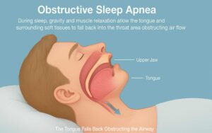
Here’s something most people don’t expect: your jaw position and your airway space are closely linked. When the lower jaw is positioned too far back, or when muscle tension pulls the jaw out of its natural alignment, it can also narrow the airway, the space air travels through when you breathe, especially during sleep.
This is why dentists increasingly look at both the jaw joint and the airway together, rather than treating TMD as an isolated bite or muscle problem. A narrowed airway can contribute to:
- Snoring and disrupted sleep
- Mouth breathing
- Daytime fatigue
- In some cases, mild to moderate sleep apnea
So, when a dentist evaluates TMD using CBCT, they’re not only checking bone structure they’re also assessing whether jaw position is quietly affecting how well you breathe at night.
How CBCT Imaging Actually Works
A CBCT scan takes just a few seconds and involves a machine rotating around your head while capturing hundreds of images. These are then reconstructed into a detailed 3D model that a dentist can rotate, zoom into, and measure precisely.
Compared to a traditional 2D X-ray or OPG, CBCT offers:
- Sub-millimeter accuracy: Allowing measurement of tiny joint changes that 2D images simply cannot show
- Full airway volume mapping: Dentists can calculate the exact cross-sectional area and volume of your airway at different points
- Bone and joint visualization from every angle: Helpful for spotting erosion, joint space, or asymmetry in the jaw joint
- Low radiation exposure: A CBCT scan typically involves a radiation dose comparable to a handful of routine dental X-rays, far lower than a medical CT scan
Because CBCT is a single scan that captures so much detail, it often reduces the need for multiple separate investigations, saving patients both time and repeat visits.
What Dentists Look for in a CBCT Airway Analysis
During a CBCT-based TMD and airway evaluation, a dentist typically assesses:
- Condyle position at rest and on mouth open position.
- Joint space symmetry comparing the left and right sides of the jaw joint
- Bony changes and signs of wear, flattening, or degeneration in the joint
- Airway volume and narrowest point identifying constriction zones in the throat area
- Tongue and soft palate position since these structures also influence airway space
By reviewing all of this together, a dentist can build a much clearer picture of whether your jaw pain, headaches, or sleep issues are connected, and design a treatment plan that addresses the actual cause, not just the symptom.
Author Bio

Foinix Digital
Foinix Digital Services believes every story has the power to inspire and connect. With a passion for creativity and meaningful storytelling, we craft content that brings ideas to life and helps brands share their voice with the world.
Recent Posts


Root Canal
April 14, 2025
Dental
May 2, 2025
Book Appointment
Why This Matters for Patients in Hyderabad
Hyderabad’s lifestyle of long commutes, high-pressure IT and business schedules, late nights, and increasing stress levels has quietly contributed to a rise in jaw clenching (bruxism) and TMD-related complaints across the city. Many patients only seek help once the pain becomes constant or starts affecting sleep and daily function.
Having access to advanced 3D imaging locally means Hyderabad residents no longer need to travel to metro hubs like Mumbai or Delhi for accurate, specialist-level TMD and airway assessment. A dental clinic equipped with in-house CBCT technology can offer same-visit imaging, faster diagnosis, and a treatment plan built on precise data rather than guesswork.
What Happens After the Scan?
Once the CBCT images are analyzed, treatment approaches for TMD can vary based on severity and cause. Common next steps may include:
- Custom neuromuscular orthotic splints to reduce clenching pressure on the joint
- Bite correction or orthodontic evaluation if jaw alignment is contributing to the issue
- Airway-focused treatment in cases where airway narrowing is significant, coordinated care with an ENT or sleep specialist may be recommended
- Lifestyle guidance including stress management and sleep posture adjustments
The goal isn’t a one-size-fits-all fix, it’s a plan built around what your specific scan reveals.
Is CBCT Right for You?
You don’t necessarily need a CBCT scan for every jaw complaint. But it’s worth discussing with your dentist if you experience:
- Jaw pain lasting more than a few weeks
- Frequent morning headaches
- Loud snoring or a partner noticing pauses in your breathing at night
- Jaw locking or difficulty opening your mouth fully
- Unexplained ear pain with no infection found
A short consultation can help determine whether imaging is the right next step for your specific symptoms.
Final Thoughts
TMD and airway health are more connected than most patients realize, and relying on outdated 2D imaging often means missing the full picture literally. With CBCT airway analysis, dentists in Hyderabad can now offer a level of diagnostic precision that was previously available only through multiple, separate investigations.
If jaw pain, clicking, or poor sleep has been part of your routine for a while, it may be time to look deeper quite literally with a 3D scan that shows exactly what’s happening beneath the surface.
FAQ's
A regular dental X-ray gives a flat, 2D image, while a CBCT scan builds a full 3D model of the jaw joint, bone, and airway. This allows dentists to measure joint space, bone changes, and airway volume with far greater accuracy than a standard X-ray allows.
Yes, Jaw position and airway space are closely linked. When the jaw sits too far back or muscle tension pulls it out of alignment, it can narrow the airway, contributing to snoring, mouth breathing, or disrupted sleep in some patients.
CBCT scans use significantly lower radiation than a medical CT scan typically comparable to a small number of routine dental X-rays. The scan itself takes under a minute, making it a quick and low-risk diagnostic option.
Common TMD signs include jaw clicking or popping, morning jaw soreness, difficulty chewing, frequent headaches, and ear pain without infection. Since these symptoms overlap with other conditions, a dental evaluation with imaging is the most reliable way to confirm TMD.
Not always, but in cases of significant airway narrowing linked to sleep-disordered breathing, dentists often coordinate care with an ENT or sleep medicine specialist to ensure the airway concern is managed alongside the jaw treatment.